AZ Logo Skip To Content
  • Tumor Types & BiomarkersTumor Types & Biomarkers

    Tumor Types & Biomarkers

    Explore clinical guidelines on why, whom and how to test actionable biomarkers in lung, breast, ovarian and prostate cancers.

    • Lung Tumor
      • EGFR
      • ERBB2 (HER2)
    • Breast Tumor
      • BRCA1/2
      • ESR1
      • HER2
      • PIK3CA/AKT1/PTEN
    • Ovarian Tumor
      • HRD
    • Prostate Tumor
      • HRRm
      • PTEN
  • Testing Solutions
  • Explore Precision MedicineTumor Types & Biomarkers

    Explore Precision Medicine

    Implement best practices in your workflow and discover how recent advances are optimizing precision medicine.

    Realizing the Potential of Precision Medicine

    Collaborate across the MDT to address health inequities and optimize testing pathways.

    Realizing the Potential of Precision Medicine

    Latest Developments in Precision Medicine

    Explore recent advances in diagnostic technologies and novel biomarkers to support precision medicine in your practice.

    Latest Developments in Precision Medicine
  • ResourcesTumor Types & Biomarkers

    Resources

    Access a range of resources you can apply in your practice.

    FAQs

    Find answers to frequently asked questions about precision medicine.

    FAQs

    Find a Testing Lab

    Search our directory of major laboratories that perform diagnostic biomarker testing.

    Find a Testing Lab

    Glossary

    Find definitions for the most commonly used terms in precision medicine.

    Glossary

    Resource Library

    Explore our library of videos and downloadable tools to help optimize your practice and inform your patients.

    Resource Library

    My Favorites

    Personalize your learning by creating a bespoke collection of precision medicine resources.

    My Favorites
  • Our Team
Subscribe Connect with us
Open MenuClose Menu

Search

  • Home Icon
  • Prostate Cancer

  • PTEN

  • PTEN

bolt

Personalize your browsing experience today.

What are you looking for today?

checking the code

PTEN Deficiency in Prostate Cancer

What is PTEN deficiency and how does its role in prostate cancer impact patients?

Explore information, recommendations for testing PTEN (phosphatase
and tensin homolog), and emerging technologies that can help inform action that may impact outcomes in metastatic prostate cancer (mPC).

PTEN Deficiency in Prostate Cancer Icon PTEN Deficiency in Prostate Cancer Icon
Reading time: 10 min
 
Jump to:
  • Clinical Relevance
  • Prevalence
  • Testing Recommendations
  • Interpreting and Reporting
  • Considerations for Your Practice
  • Helpful Resources
PTEN Biomarker in Prostate Cancer Icon

PTEN Is a Prognostic Biomarker With OS and PFS Implications1,2

Early testing for PTEN deficiency is critical1,2

PTEN deficiency is an independent predictor for shorter OS and PFS
(compared with PTEN proficiency in patients with de novo mHSPC)

PTEN Deficiency Leads to Shorter OS3
Median OS: 43.8 vs 80.2 months (P<0.001)

43.8 mo

80.2 mo

Adjusted HR: 1.67
(1.14–2.43, P=0.008)

PTEN altered

PTEN non-altered

PTEN alterations were associated with a 36.4-month difference in OS, a significantly shorter span vs non-altered PTEN.

HR, hazard ratio; mHSPC, metastatic hormone-sensitive prostate cancer; OS, overall survival.

PTEN deficiency is an independent prognostic factor for worse progression-free survival2,3

  • A real-world study found that PFS was shorter in PTEN-deficient mPC patients2
  • Patients with non-altered PTEN experienced 6.5 months longer PFS2

mPFS in mPC by PTEN status2
mPFS: 31.0 vs 37.5 months (P=0.019)

PTEN
alterations

31.0

PTEN
non-altered

37.5

0

10

20

30

40

Months

HR: 0.80 (0.66 - 0.96)

HR, hazard ratio; mPC, metastatic prostate cancer; mPFS, median progression-free survival; PFS, progression-free survival.

PTEN Deficiency Impact on Prostate Cancer Patients Checklist

PTEN deficiency negatively impacts outcomes in patients with prostate cancer and is emerging as an important biomarker in de novo mHSPC1,2

PTEN Regulates Cell Growth and Tumor Suppression

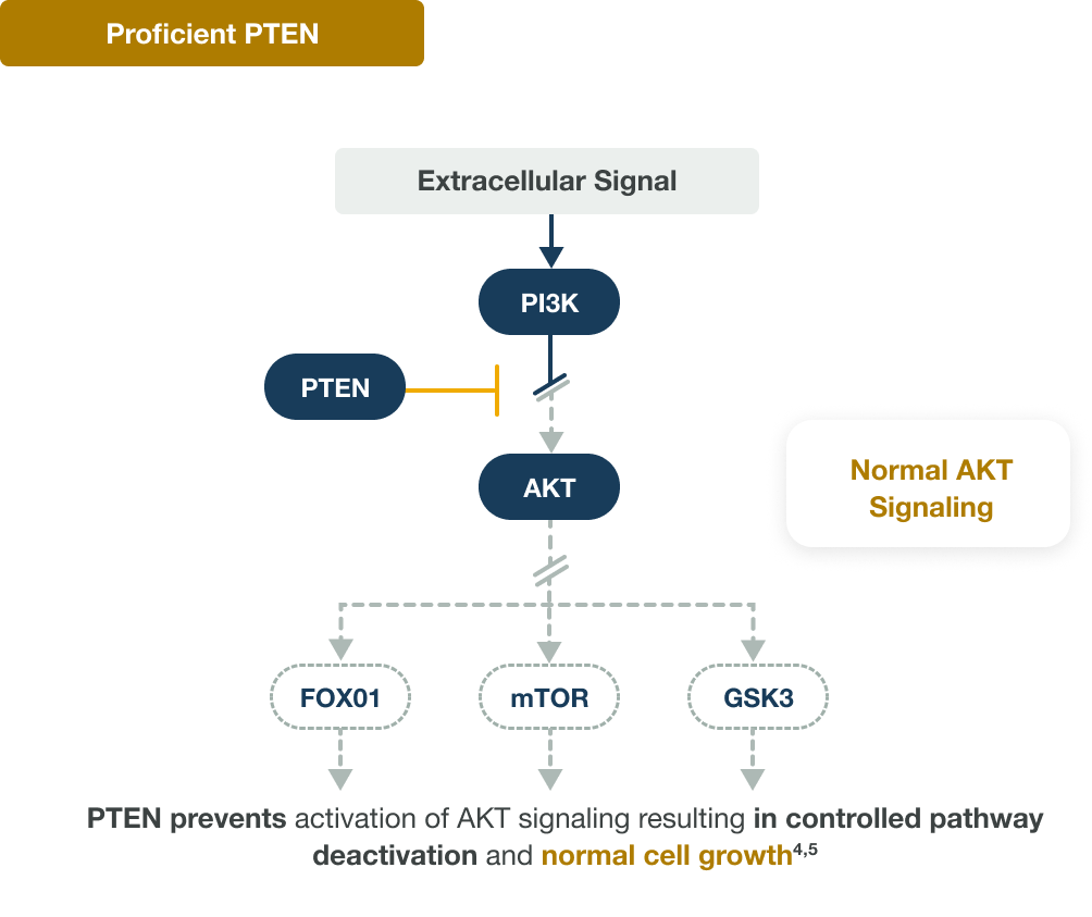
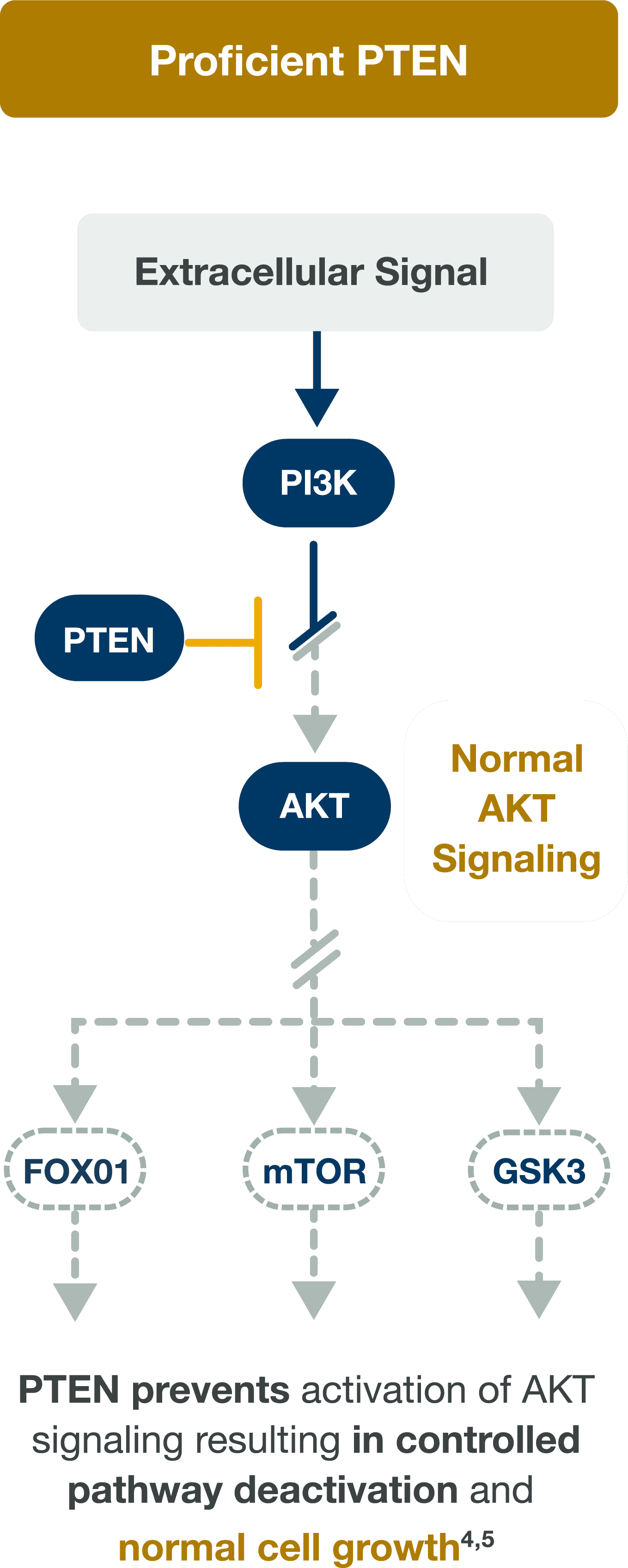
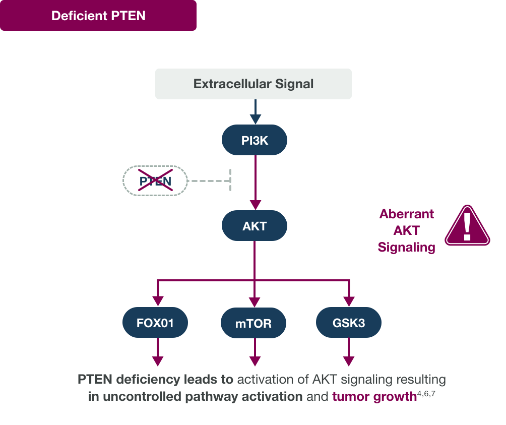
PTEN regulates cell growth and suppression of tumors4

Proficient PTEN and Normal AKT Signaling Pathway Diagram Proficient PTEN and Normal AKT Signaling Pathway Diagram
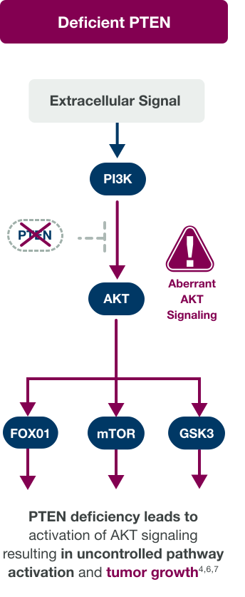
PTEN Deficiency and Aberrant AKT Signaling Pathway Diagram PTEN Deficiency and Aberrant AKT Signaling Pathway Diagram

AKT, AKR strain transforming; FOX01, Forkhead box 01; GSK3, glycogen synthase kinase-3; mTOR, mammalian target of rapamycin; PI3K, phosphoinositide 3-kinase; PTEN, phosphatase and tensin homolog.

PTEN is a Tumor Suppressor Checklist Icon

PTEN is a tumor suppressor that negatively regulates the PI3K–AKT pathway; intact PTEN maintains controlled cell growth, while PTEN loss leads to unchecked AKT signaling and tumor progression.4-7

PTEN Prevalence:
Nearly 25% of mHSPC Patients Are PTEN Deficient8

24%

PTEN deficient

(258/1095)

76%

PTEN proficient

(837/1095)

Multi-institutional cohort of surgically treated patients with PC and known ETS-related gene (ERG) status. A total of 1275 primary prostate tumors from the Canary Foundation retrospective PC tissue microarray cohort were used in this study. The tissue microarray included samples from men with recurrent PC, nonrecurrent PC, and unknown outcomes because of inadequate follow-up
time (ie, censoring).8

IHC Biomarker Testing for PTEN-Deficient Patients

IHC, in Addition to NGS, Enhances Identification of All PTEN-Deficient Patients1,9

Why IHC biomarker testing should be routine at mPC diagnosis1,2

PTEN deficiency can lead to significantly faster time to progression and worse outcomes; therefore, it is critical to ensure all patients with PTEN deficiency are identified.

Homozygous deletions, followed by truncations, are the most common PTEN alterations in prostate cancer10

Fusion

Missense

Splice

Homozygous deletion

mHSPC

Truncating mutation

~90% of PC PTEN alterations8

Genomic PTEN deletions and truncating mutations may result in loss of detectable protein that can be
assessed by IHC11

IHC is another testing method for PTEN deficiency in mPC due to the potential that NGS alone may fail to identify nearly 1 in 51,9,12

PTEN loss can occur via various nongenetic mechanisms13

NGS (next-generation sequencing) is a well-validated and convenient assay for identification of genomic alterations in a large panel of biomarkers.12

  • While NGS can detect genomic PTEN alterations, IHC (immunohistochemistry) should be used to detect PTEN protein deficiency14,15
  • Pre- and post-transcriptional factors also influence PTEN expression and would not be revealed through NGS alone13,15,16
  • IHC requires less tumor tissue vs NGS15

NGS limitations underscore the importance of IHC testing1,9

PTEN-deficient PC detected by NGS

PTEN-deficient PC missed by NGS but detectable with IHC

Nearly 1 in 5 PTEN-deficient patients are missed by NGS alone1,9

PTEN loss status was defined as a minimum of 50% of the specimen's tumor area with no detectable PTEN staining.

PTEN-Deficient Tumors Checklist Icon

Relying solely on NGS may overlook PTEN-deficient tumors in mPC1,9

IHC and NGS for Identifying PTEN-Deficient Patients Icon

IHC Testing Identifies PTEN Deficiency Caused by Genomic Alterations and Factors Beyond the Genome11,13,15,17

IHC testing utilizes antibodies specific for an antigen of interest to evaluate the presence of that antigen in a tissue sample.

IHC is commonly used to assess patient samples for changes in PTEN protein concentrations15

Advantages of IHC for PTEN testing

Biomarker Monitoring Without Disrupting Tissue Icon

Monitoring of biomarker without disrupting the architecture of the tissue—especially relevant in tumors with high intratumoral heterogeneity12

Sensitive and Specific Testing Approach Icon

Simple yet sensitive and specific approach that avoids many of the technical limitations of NGS15,18

IHC Detects PTEN Protein Levels Icon

IHC detects PTEN protein levels instead of relying on DNA sequencing, revealing PTEN loss that may be undetectable by FISH/NGS/​ctDNA14,16

Cost-Effective Prostate Cancer Screening Icon

Less expensive and less time-consuming than FISH and NGS for the routine screening of prostate tumor specimens14,15

Requires Less Tumor Tissue Icon

Requires less tumor tissue than NGS, which is appropriate in PC due to the limited size of biopsies15

Systemic PTEN IHC Analysis in mPC Icon

Systemic PTEN IHC analysis could be easily and cost-effectively implemented in the diagnostic workup of patients with mPC12

ctDNA, circulating tumor DNA; FISH, fluorescence in situ hybridization.

IHC-based analyses are best to determine the level of PTEN expression in histological samples.17

Checklist Icon 3

IHC is the only prospectively validated test and is considered the gold standard for testing PTEN deficiency.12

Interpreting and Reporting PTEN IHC 

PTEN status evaluated by IHC and NGS has different clinical implications. IHC allows for easy visualization of PTEN protein expression levels and evaluation for PTEN deficiency. NGS can provide insight into genomic alterations.15

To dive deeper into PTEN IHC interpretation and view clinical slides, click here: pioneerhubprostate.com

Visually Accessible Analysis
With IHC20

PTEN proficient

Presence of PTEN-specific
antibody staining

Stained

PTEN Proficient IHC Staining

PTEN deficient

Presence of PTEN-specific
antibody staining

Absence of PTEN-specific
antibody staining

Unstained

PTEN Deficient IHC Staining

Simple and Sensitive Clinical Interpretation

PTEN deficiency has been defined in literature as ≥90% of viable malignant cells with no specific cytoplasmic staining.19-21

  • Accurate reporting should clearly state PTEN deficiency and percentage of staining loss

A PTEN IHC Assay Scoring Algorithm19-21

Clinical interpretation:
PTEN status

Staining pattern

Deficienta

≥90% viable malignant cells with no specific cytoplasmic
staining

Proficienta

>10% viable malignant cells with any specific cytoplasmic stain intensity

aProstatic adenocarcinoma tissue samples are assessed for PTEN deficiency in the presence of acceptable controls. Nuclear staining of viable malignant cells must be disregarded.

aProstatic adenocarcinoma tissue samples are assessed for PTEN deficiency in the presence of acceptable controls. Nuclear staining of viable malignant cells must be disregarded.

PTEN NGS testing quantifies mutations, copy-number losses, and loss of heterozygosity across the gene. Pathogenic variants, single-copy deletions, or deep (biallelic) losses indicate disrupted PTEN function. This provides a comprehensive view of how the PI3K/AKT pathway is genomically activated.4,19,22

Key Takeaways for PTEN Testing in Prostate Cancer Icon

Considerations for Your Practice

Key Takeaways for PTEN Testing in Prostate Cancer

  • Patients who are PTEN deficient have worse OS, PFS, and general outcomes of standard of care treatment than patients who are PTEN proficient1,2
  • PTEN plays an important role in cell growth by regulating the AKT pathway and ensuring normal cell division23
  • IHC testing may identify patients with PTEN deficiency who are missed with NGS testing alone. Testing should be conducted with IHC to confirm PTEN status1,9
  • PTEN deficiency is defined as ≥90% of viable malignant cells with no specific cytoplasmic staining19-21

OS, overall survival; PFS, progression-free survival.

Pathologist Reviewing Lab Sample for PTEN Testing ImagePathologist Reviewing Lab Sample for PTEN Testing Image

Ensure PTEN deficiency is clearly reported with the percentage of protein loss. Collaboration between pathologists, labs, and MDT (multidisciplinary team)
on PTEN testing, ordering, and reporting may help
improve patient care.

  • View educational resources that can help facilitate conversations between you and your MDT colleagues treating metastatic prostate cancer at pioneerhubprostate.com

Helpful Resources

Check out these featured resources on PTEN biomarker testing in metastatic prostate cancer. 

Playlist icon

Add your preferred resources to My Favorites—a personalized list of resources to download and share with your peers or patients.

View My Favorites

Title of video goes here lorem ipsum ae prae rem quunt prepuda ntiurep recest
0:00 MIN
Title of video goes here lorem ipsum ae prae rem quunt prepuda ntiurep recest

Description of vide goes here lorem ipsum cae prae rem quunt prepuda ntiurep recest hitissum aut assunto odiciis etum quam re, sam re conet reicae netur atemo exerum expligendant expelendel ipsum vercips apidit fugia ex expe et lacepuda con nullum iur sinullab inctae consequi aut

Title of video goes here lorem ipsum ae prae rem quunt prepuda ntiurep recest
0:00 MIN
Title of video goes here lorem ipsum ae prae rem quunt prepuda ntiurep recest

Description of vide goes here lorem ipsum cae prae rem quunt prepuda ntiurep recest hitissum aut assunto odiciis etum quam re, sam re conet reicae netur atemo exerum expligendant expelendel ipsum vercips apidit fugia ex expe et lacepuda con nullum iur sinullab inctae consequi aut

Title of video goes here lorem ipsum ae prae rem quunt prepuda ntiurep recest
0:00 MIN
Title of video goes here lorem ipsum ae prae rem quunt prepuda ntiurep recest

Description of vide goes here lorem ipsum cae prae rem quunt prepuda ntiurep recest hitissum aut assunto odiciis etum quam re, sam re conet reicae netur atemo exerum expligendant expelendel ipsum vercips apidit fugia ex expe et lacepuda con nullum iur sinullab inctae consequi aut

View more resources
Email icon

Subscribe to Emails

Subscribe and stay up to date with biomarker testing
in metastatic prostate cancer.

Subscribe

Chat Icon

Connect With Us

Connect with our Precision Medicine Team for actionable
support PTEN-deficiency testing with IHC.

Connect with us

  1. de Bono J, et al. 2021. Presented at ASCO-GU Cancers Symposium 2021. Abstract #13.
  2. Thapa B, et al. Ann Oncol. 2024;35:S991.
  3. Zhang J-Y, et al. Asian J Androl. 2022;24:50–55.
  4. Jamaspishvili T, et al. Nat Rev Urol. 2018;15:222–234.
  5. Brown JS, Banerji U. Pharmacol Ther. 2017;172:101–115.
  6. Marques RB, et al. Eur Urol. 2015;67(6):1177–1185.
  7. Ferraldeschi R, et al. Eur Urol. 2015;67(4):795–802.
  8. Stopsack KH et al. Clin Oncol Res. 2020;26:3230-3238.
  9. Sweeney C, et al. Lancet. 2021;398:131–142.
  10. Gilson C, et al. JCO Precis Oncol. 2020;4:882–897.
  11. Pulido R, et al. Cold Spring Harb Perspect Med. 2019;9:a036293.
  12. Giunta EF, et al. Cancers (Basel). 2021;13(19):4771.
  13. Leslie NR, et al. Trends Pharmacol Sci. 2011;32(3):131–140.
  14. Lotan TL, et al. Mod Pathol. 2016;29(8):904-914.
  15. Tsao MS, Yatabe Y. J Thorac Oncol. 2019;14:2035–2038.
  16. Wise HM, et al. Clin Sci (Lond). 2017;131:197–210.
  17. Castillo-Martin M, et al. Methods Mol Biol. 2016;1388:23–37.
  18. Shah RB, et al. Prostate. 2019;79:1267–1273.
  19. Lotan TL, et al. Clin Cancer Res. 2011;17(20):6563–6573.
  20. Lotan TL, et al. Oncotarget. 2017;8(39):65566–65576.
  21. Fizazi K, et al. Ann Oncol. 2026;37(1):53-68.
  22. Turnham, DJ et al. Cells. 2020;9:2342.
  23. Tortorella E, et al. Int J Mol Sci. 2023;24:2046.
AstraZenecaYoutube

This information is intended for US healthcare professionals only.

Connect With Us Subscribe to Emails Sitemap
Privacy Notice Legal StatementAstraZeneca US Corporate Site Contact Us
Cookie Policy Cookie Notice Consumer Health Data Privacy Notice Your Privacy ChoicesYour Privacy ChoicesYour Privacy Choices

©2026 AstraZeneca. All rights reserved. US-107047 Last Updated 2/26

Share Resource

Share your precision medicine resource(s) via social media or email.

Share to Apps

{{$.copyrighttext}}

©2024 AstraZeneca. All rights reserved. US-86234 Last Updated 11/24

  • Share via  LinkedIn
  • Share via  Facebook
  • Share via  X
  • Share via  Doximity
Share via Email
To add multiple email addresses, please separate them with a comma (e.g. email1@domain.com, email2@domain.com)

errorMessage

requestToSignInContent

Welcome to

AZPrecisionMed.com

This website is intended to help healthcare professionals practicing in the US find and access scientifically balanced and evidence-based information and resources on oncology biomarker testing in precision medicine.

The dissemination of this information may be subject to different medical and regulatory requirements in other countries.

This information is provided for educational purposes only. It is not intended for use in the diagnosis or treatment of individual patients.

Are you a healthcare professional in the United States?

No, take me to AstraZeneca-us.com

What is an AstraZeneca authorised person?

This website is only intended for healthcare professionals practicing in the United States and authorized AstraZeneca company persons. US Headquarters personnel, US Field Diagnostics and US Field Medical personnel are authorized to browse and search the database for their own background knowledge and insight. This website is not intended for use by US Field Sales personnel. None of the content on this site should be directly linked or distributed without approval.

©2024 AstraZeneca. All rights reserved. US-86234 Last Updated 11/24

  • Share via  LinkedIn
  • Share via  Facebook
  • Share via  X
  • Share via  Doximity
Share via Email
To add multiple email addresses, please separate them with a comma (e.g. email1@domain.com, email2@domain.com)

errorMessage

requestToSignInContent

Share Resource

Share your precision medicine resource(s) via social media or email.

Share to Apps

©2024 AstraZeneca. All rights reserved. US-86234 Last Updated 11/24