AZ Logo Skip To Content
  • Tumor Types & BiomarkersTumor Types & Biomarkers

    Tumor Types & Biomarkers

    Explore clinical guidelines on why, whom and how to test actionable biomarkers in lung, breast, ovarian and prostate cancers.

    • Lung Tumor
      • EGFR
      • ERBB2 (HER2)
    • Breast Tumor
      • BRCA1/2
      • ESR1
      • HER2
      • PIK3CA/AKT1/PTEN
    • Ovarian Tumor
      • HRD
    • Prostate Tumor
      • HRRm
      • PTEN
  • Testing Solutions
  • Explore Precision MedicineTumor Types & Biomarkers

    Explore Precision Medicine

    Implement best practices in your workflow and discover how recent advances are optimizing precision medicine.

    Realizing the Potential of Precision Medicine

    Collaborate across the MDT to address health inequities and optimize testing pathways.

    Realizing the Potential of Precision Medicine

    Latest Developments in Precision Medicine

    Explore recent advances in diagnostic technologies and novel biomarkers to support precision medicine in your practice.

    Latest Developments in Precision Medicine
  • ResourcesTumor Types & Biomarkers

    Resources

    Access a range of resources you can apply in your practice.

    FAQs

    Find answers to frequently asked questions about precision medicine.

    FAQs

    Find a Testing Lab

    Search our directory of major laboratories that perform diagnostic biomarker testing.

    Find a Testing Lab

    Glossary

    Find definitions for the most commonly used terms in precision medicine.

    Glossary

    Resource Library

    Explore our library of videos and downloadable tools to help optimize your practice and inform your patients.

    Resource Library

    My Favorites

    Personalize your learning by creating a bespoke collection of precision medicine resources.

    My Favorites
  • Our Team
Subscribe Connect with us
Open MenuClose Menu

Search

  • Home Icon
  • Breast cancer

  • HER2

  • HER2

bolt

Personalize your browsing experience today.

What are you looking for today?

checking the code

HER2 Expression in Breast Cancer

Explore guideline recommendations and actionable best practices for HER2 IHC testing in breast cancer.

Header icon for the HER2 Expression in Breast Cancer webpage Header icon for the HER2 Expression in Breast Cancer webpage
Reading time: 22 min
 
Jump to:
  • HER2 Expression
  • Clinical Relevance
  • Prevalence
  • Diagnostic Assays
  • Interpreting & Reporting
  • HER2-low Expression
  • CAP HER2 IHC Reporting
  • Considerations for Your Practice
  • Helpful Resources

Scoring HER2 Expression Across the Full Spectrum Helps to Inform Treatment Decisions1,2

Knowing human epidermal growth factor receptor 2 (HER2) status in breast cancer guides care options and informs eligibility for targeted therapies, which is why the NCCN Clinical Practice Guidelines In Oncology (NCCN Guidelines®) recommend testing for this biomarker in all patients with breast cancer.1

Checklist icon small

Since 2007, the American Society of Clinical Oncology (ASCO) and College of American Pathologists (CAP) have provided guidance for HER2 immunohistochemistry (IHC) testing.2 These clinical practice guidelines are periodically updated to meet changing practice needs and incorporate new evidence.

Previously, clinical classification of HER2 status was binary, with assays and best practices optimized to identify HER2-positive (IHC 3+ or IHC 2+/in situ hybridization [ISH]+) breast tumors vs HER2-negative.3 This changed with the 2023 ASCO/CAP guideline update that offers additional guidance around reporting HER2 IHC 1+, IHC 2+/ISH- results, and best practices for differentiating IHC 1+ and IHC 0 tumors.2

ASCO, American Society of Clinical Oncology; CAP, College of American Pathologists; HER2, human epidermal growth factor receptor 2; IHC, immunohistochemistry; ISH, in situ hybridization; NCCN, National Comprehensive Cancer Network.

Checklist icon light

HER2 Expression May Inform Prognostic and Predictive Outcomes4,5

The pathologist's role in HER2 status assessment is critical for targeted treatment

HER2 is overexpressed in approximately 15% of primary breast cancers.4 HER2 is both prognostic and predictive in breast cancer.5 When activated, it causes tumor cell proliferation and drives disease progression.5 Breast cancers are classified by hormone receptor (HR) status (positive or negative; HR+/-) and HER2 status (positive or negative).6

Checklist icon small

The development of HER2-directed therapies has been practice-changing and has significantly improved overall survival in the HER2-positive breast cancer population.5,7 Consequently, testing for this biomarker has become a routine part of the diagnostic work-up for patients with breast cancer.5

HER2, human epidermal growth factor receptor 2; HR, hormone receptor.

DNA and patient icon

Around 6 in 10 Patients Categorized as HER2-Negative Have Discernible Low Levels of HER2 Expression4,8,9

Figure showing that, of all breast cancers, ~85% are HER2-negative and ~15% are HER2-positive. Of patients categorised as HER2-negative, 6 in 10 have discernible low levels of HER2 expression ^4,8,9

HER2-negative

~85%

HER2-positive

~15%

IHC 0

IHC 1+

IHC 2+/

ISH-

IHC 2+/

ISH +

IHC 3+

HER2-low

~60% of HER2-negative

 Figure showing that, of all breast cancers, ~85% are HER2-negative and ~15% are HER2-positive. Of patients categorised as HER2-negative, 6 in 10 have discernible low levels of HER2 expression  ^4,8,9

HER2-negative

~85%

HER2-positive

~15%

IHC 0

IHC 1+

IHC 2+/
ISH-

IHC 2+/
ISH+

IHC 3+

HER2-low

~60% of HER2-negative

Adapted from Tarantino P, et al.; Schettini F, et al. and Rakha EA, et al.

Discrete scoring across the full spectrum of HER2 expression further differentiates each patient2,4

As clinical research evolves, it is becoming increasingly relevant to evaluate and differentiate each case of breast cancer with a discrete IHC score.2,4 This is due to the clinical significance of HER2 extending beyond the binary classification of positive or negative.4 A subset of HER2-negative expression has now been defined as HER2-low (IHC 1+ or IHC 2+/ISH-).8,10 Approximately 60% of patients with breast cancer who are categorized as HER2-negative have discernible low levels of HER2 expression.8 Ongoing research is investigating whether breast cancers scored as HER2 IHC 1+ and IHC 2+/ISH- show biological differences from those scored as IHC 0.4,8

Biomarker DNA icon

The CAP Breast Biomarker Reporting template states that HER2 IHC 1+ and IHC 2+/ISH- may be reported as HER2-low.11

CAP, College of American Pathologists; HER2, human epidermal growth factor receptor 2; IHC, immunohistochemistry; ISH, in situ hybridization.

Laboratory microscope light icon

There Are Four FDA-Approved Diagnostic Assays for Measuring HER2 Alterations12

ASCO/CAP guidelines recommend the use of FDA-approved assays for HER2 testing in breast cancer.13

Assay name
(alphabetical order)
Bond™ Oracle™ HER2 IHC System HercepTest™ InSite™ HER-2/neu PATHWAY anti-HER2/neu
Platform BOND-MAX14 Dako Automated Link Platform16 BioGenex i6000™ Automated Staining System or the OptiMax® Plus Consolidated Staining System17 BenchMark ULTRA18
Antibody clone/type CB1114,15
Mouse monoclonal antibody
RpAb16 Rabbit polyclonal antibody CB1117
Mouse monoclonal antibody
4B518 Rabbit monoclonal antibody
Manufacturer Leica Biosystems14,15 Agilent/Dako16 BioGenix17 Roche/Ventana18

See a list of major testing labs in your area.

Find a Testing Lab
Hand of scientist holding petri dish

This information is intended as educational and is not intended as a complete list of available testing options. AstraZeneca is not responsible for any test provider and does not endorse any particular diagnostic test. The accuracy and results of diagnostic tests vary, and AstraZeneca shall have no liability arising from such testing. Information provided herein should in no way be considered a guarantee of coverage, reimbursement, or patient assistance. Providers should contact third-party laboratories for information on their patient assistance programs. While diagnostic testing may assist providers in identifying appropriate treatment for patients, the decision and action should be decided by a provider in consultation with the patient. All products are trademarks of their respective holders, all rights reserved.

ASCO, American Society of Clinical Oncology; CAP, College of American Pathologists; FDA, US Food and Drug Administration; HER2, human epidermal growth factor receptor 2.

Results icon

How HER2 Results Are Interpreted and Reported Can Have Implications for Patient Care

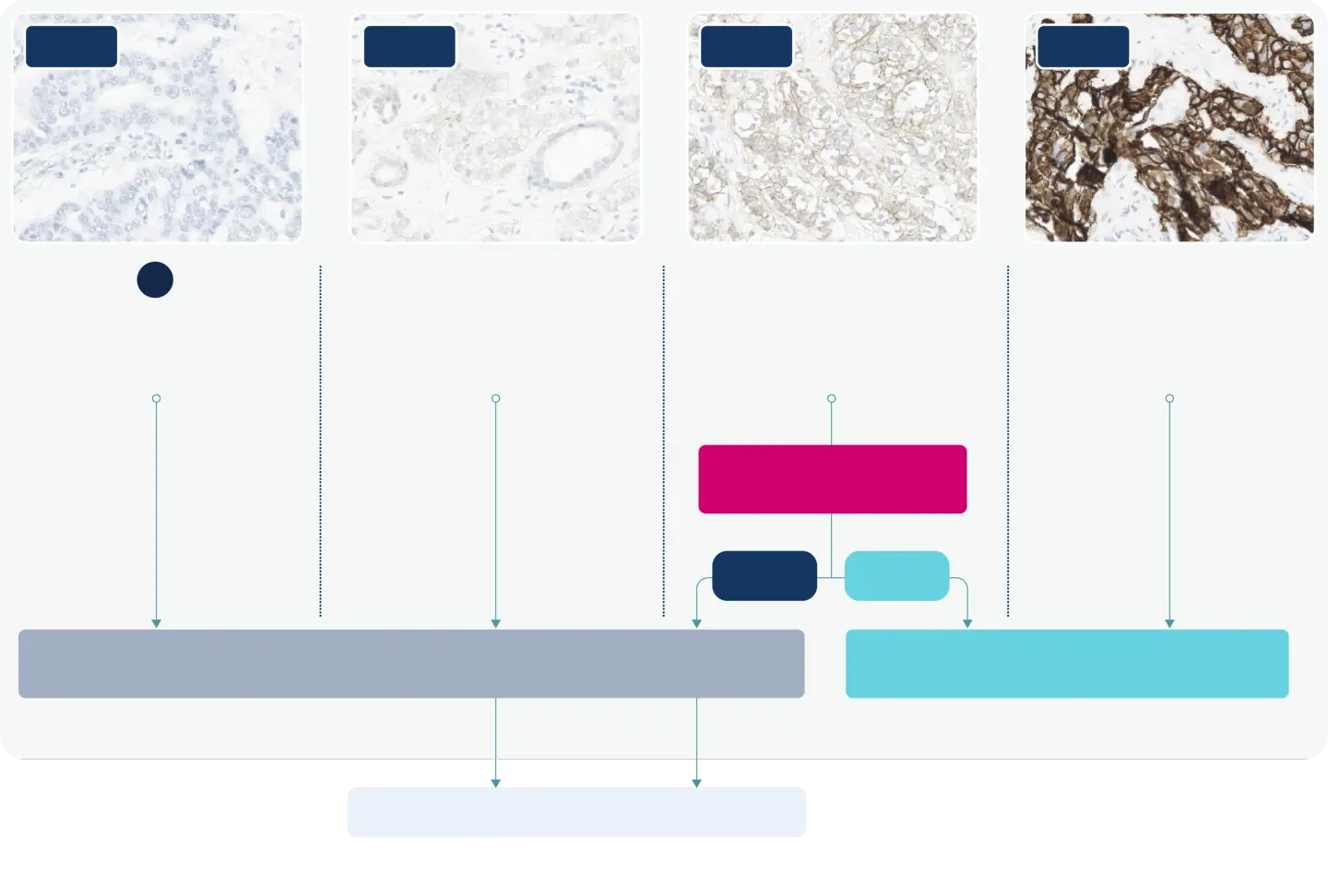
An alternative ASCO/CAP scoring algorithm to reflect the complete range of HER2 expression1,2

Four slides showing increasing IHC scale from 0 to 3+ and the type of immunohistochemistry staining that may be obtained including: no or incomplete staining in ≤ 10% tumor cells for IHC 0; incomplete membrane staining in >10% tumor cells for IHC 1+; complete membrane staining in >10% tumor cells for IHC 2+; and complete circumferential membrane staining in >10% tumor cells.

IHC 0

IHC 1+

IHC 2+

IHC 3+

No membrane staining

OR

Incomplete membrane staining Faint/barely perceptible ≤10% tumor cells

Incomplete membrane staining
Faint/barely perceptible >10% tumor cells

Complete membrane staining 

Weak-moderate >10% tumor cells

Complete circumferential membrane staining Intense >10% tumor cells

Equivocal

Reflex ISH Negative

Reflex ISH Positive

HER2-negative

HER2-positive

Additional research required

May benefit from targeting HER2

HER2-low

May benefit from targeting HER2

Images contributed from HER2Know.com.19 Figure adapted from 2023 ASCO/CAP guidelines HER2 testing algorithm.2

Use controls with a wide range of HER2 expression including IHC 1+ staining to help ensure that the assay has an appropriate limit of detection.2

HER2, human epidermal growth factor receptor 2; IHC, immunohistochemistry; ISH, in situ hybridization.

Investigate DNA icon

Scoring HER2-Low Expression May Be Clinically Relevant for Patients With Breast Cancer20

Identifying HER2-low expression relies on two important factors: the consistent distinguishing between IHC 0 and IHC 1+ and the seamless communication of discrete HER2 IHC scores between pathologists and oncologists.2 Differentiating these staining patterns can be difficult; however, reliable reporting of scores at the low end of the spectrum may be clinically meaningful for patients with breast cancer.1

HER2, human epidermal growth factor receptor 2; IHC, immunohistochemistry.

Checklist icon light

Interpret and Classify HER2 Expression Status Using ASCO/CAP Testing Guidelines2

The 2023 ASCO/CAP guideline update provides recommendations for interpreting and reporting low expression of HER2.2 The ASCO/CAP guidelines define HER2 IHC 0 as either no staining is observed or membrane staining that is incomplete and is faint/barely perceptible and in ≤10% of tumor cells.2,21 Meanwhile, IHC 1+ is defined as incomplete membrane staining that is faint/barely perceptible and in >10% of tumor cells.2

Samples on the border of IHC 0 and 1+ can be difficult to interpret

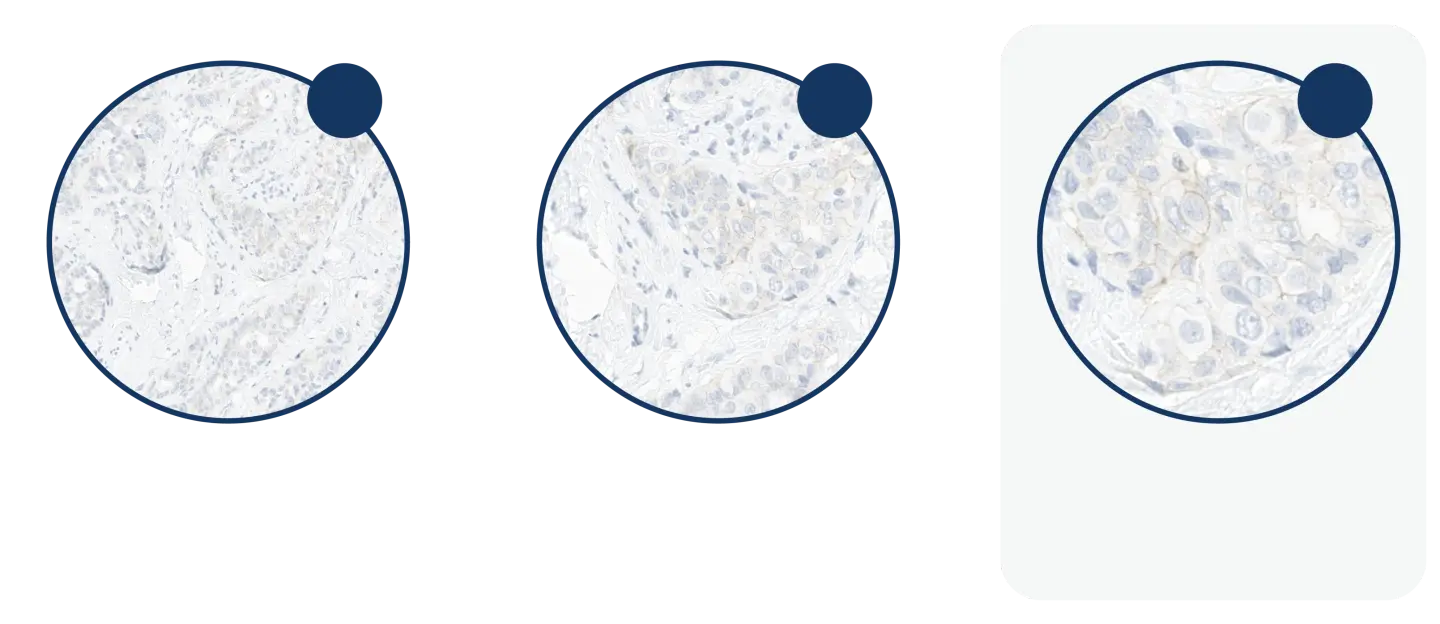
Microscopic images increasing from 10x to 40x, illustrating the magnification required to discriminate between IHC 0, 1+ and 2+.

IHC 0

IHC 1+

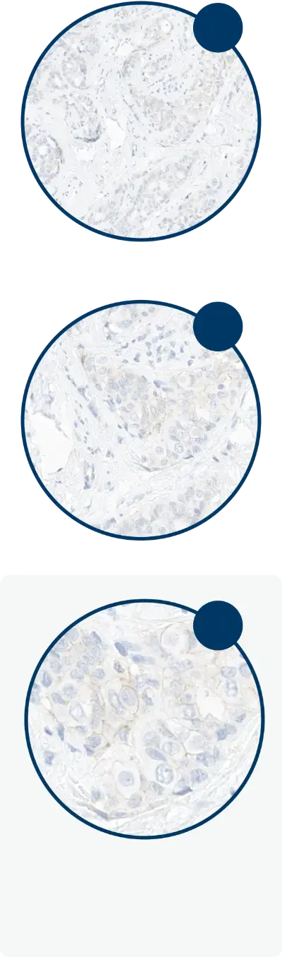
Cases can be examined at 40x or higher power magnification to discriminate between IHC 0 and 1+.2

Three microscopic images increasing from 10x to 40x, illustrating the magnification required to discriminate between IHC 0, 1+ and 2+.

10x

20x

40x

Up to 40x magnification may be required to fully discriminate among IHC 0, 1+, and 2+

Three microscopic images increasing from 10x to 40x, illustrating the magnification required to discriminate between IHC 0, 1+ and 2+.

10x

20x

40x

Up to 40x magnification

may be required to fully discriminate among IHC 0, 1+, and 2+

Images contributed from HER2Know.com.19

Tips for differentiating between IHC 0 and IHC 1+2:

Checklist icon small
  • Examine HER2 IHC-stained slides using standardized ASCO/CAP guidelines scoring criteria
Magnification x40 icon
  • Examine HER2 IHC at high power magnification (40x) when discriminating 0 from IHC 1+ staining
Healthcare professional icon
  • Consider a second pathologist review when results are close to the 0 versus IHC 1+ interpretive threshold (>10% of cells with incomplete membrane staining that is faint/barely perceptible)
Controls icon
  • Use controls with a range of protein expression (including 1+) to help ensure the assay has an appropriate limit of detection
Tissue sample icon
  • Give careful attention to preanalytical conditions of breast cancer tissue samples from both primary and metastatic sites

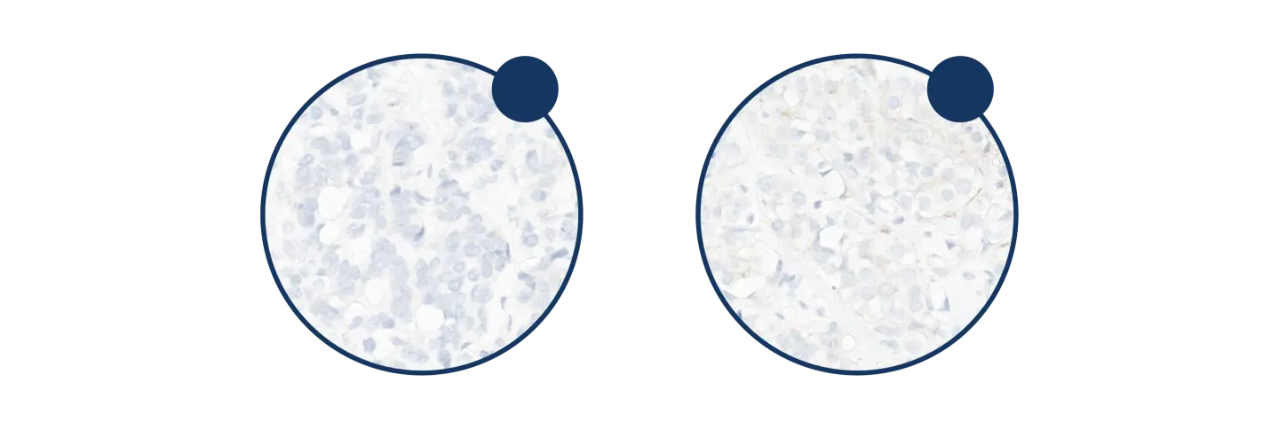
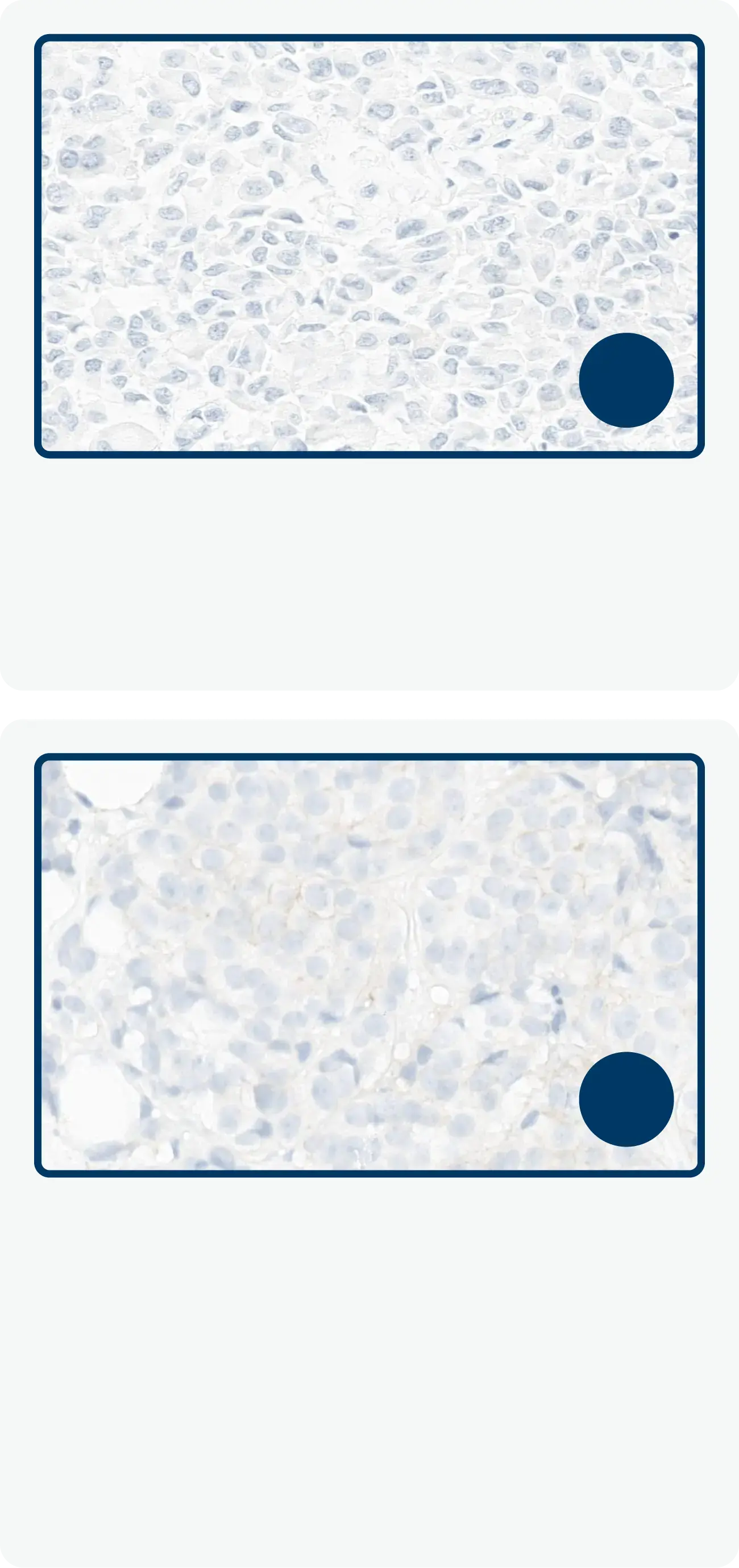
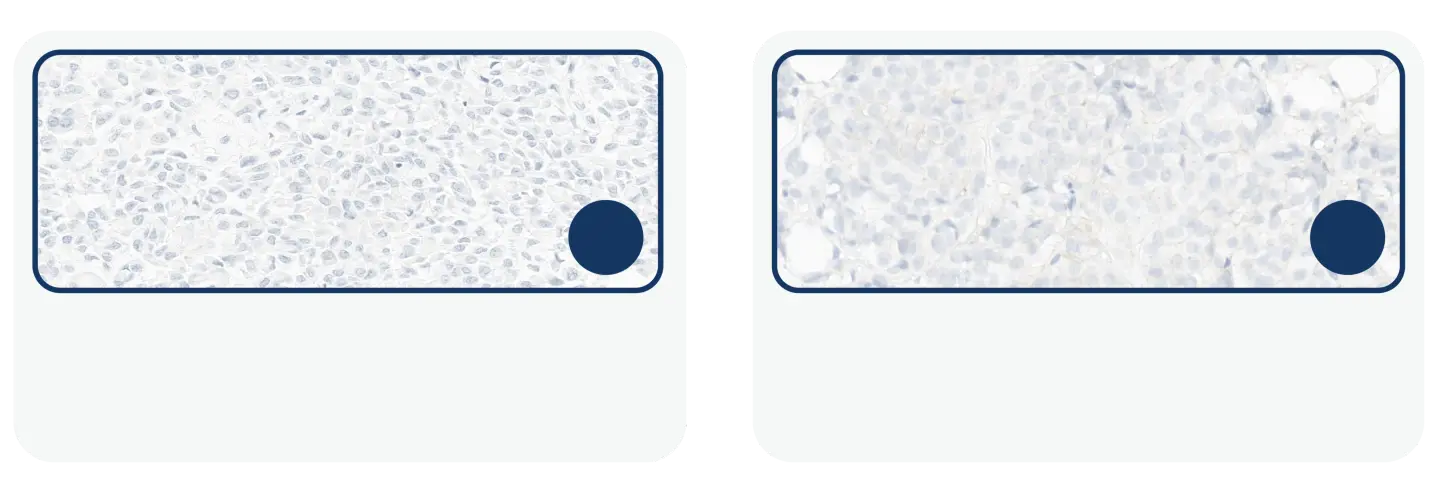
High power magnification (40x) may also help differentiate the distinct types of HER2 IHC 0 results (with and without staining):

HER2 IHC

40x

HER2 IHC 0

No staining is observed

40x

HER2 IHC 0

Membrane staining that is incomplete and faint/barely perceptible, and in ≤10% of tumor cells

HER2 IHC

40x

40x

HER2 IHC 0

No staining is observed

HER2 IHC 0

Membrane staining that is incomplete and faint/barely perceptible, and in ≤10% of tumor cells

Images contributed from HER2Know.com.19

ASCO, American Society of Clinical Oncology; CAP, College of American Pathologists; HER2, human epidermal growth factor receptor 2; IHC, immunohistochemistry.

Guidelines icon

Suboptimal Preanalytic Factors Can Prevent Consistent Interpretation of HER2 Expression Levels in Breast Cancer4

Sample collection

  • Core needle biopsies may not account for heterogeneity, and are prone to edge effects and crush artifacts2,22
  • If the initial HER2 test result in a core needle biopsy is negative or equivocal, a new HER2 test may be ordered on the excision specimen2
  • Scan the entire tissue section to ensure scoring is carried out on well-preserved and well-stained areas2

HER2 signal

  • Time lapse decreases HER2 signal intensities2,22

Time to fixation

  • Delays in time from tissue acquisition to fixation (i.e., cold ischemic time) can compromise validity of HER2 test results2,22

Fixation consistency

  • Fixation variables may artificially drive down the detection of protein expression2,22
  • Keep time to fixation as short as possible; specimens should be sliced into 5–10 mm intervals and fixed in 10% NBF for 6–72 hours2

Artifacts

  • An awareness of potential artifacts is important to avoid over-interpretation of aberrant staining22

Quality assurance

  • Validate HER2 IHC assays according to the most recent ASCO/CAP guidelines2
  • Request additional tests if HER2 IHC cannot be determined due to lack of appropriate controls, evidence of inadequate specimen handing, artifacts, or analytic testing failure2

Find best practices to address sample limitations.

Browse Testing Solutions
Hand of scientist holding petri dish

ASCO, American Society of Clinical Oncology; CAP, College of American Pathologists; HER2, human epidermal growth factor receptor 2; IHC, immunohistochemistry; NBF, neutral buffered formalin.

Guideline Recommendations Can Provide Consistency When Interpreting Challenging HER2 IHC Results2

Adhering to 2023 ASCO/CAP guideline recommendations can help ensure consistency when interpreting HER2 expression results by IHC.2

Find out more about guideline recommendations for HER2 testing.

Browse Latest Developments
Microscopic slide showing stained cells that are positive for HER2 using immunohistochemistry (IHC)

Solving common HER2 IHC interpretation challenges

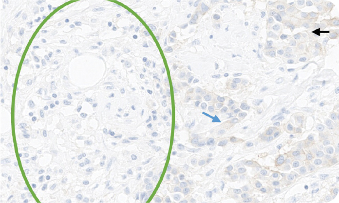
Microscopic image displaying different types of unusual staining patterns during HER2 interpretation.
Distinguishing membrane staining from cytoplasmic, basal only, and nuclear staining can make HER2 IHC interpretation challenging.23
IHC 1+ (20x original magnification). About 15% of the tumor cells form a distinct focus displaying a faint and incomplete membranous staining (black arrow), while the rest of the tumor is negative (green circle). Some of the tumor cells display a non-specific, faint cytoplasmic blush (blue arrow) which does not affect evaluation.
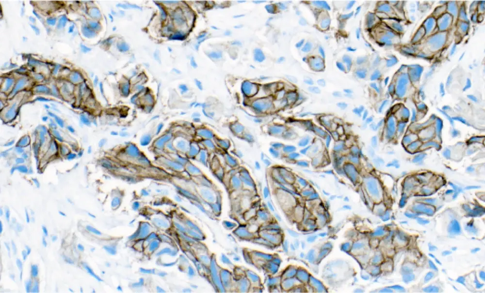
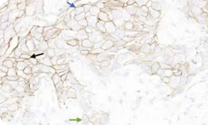
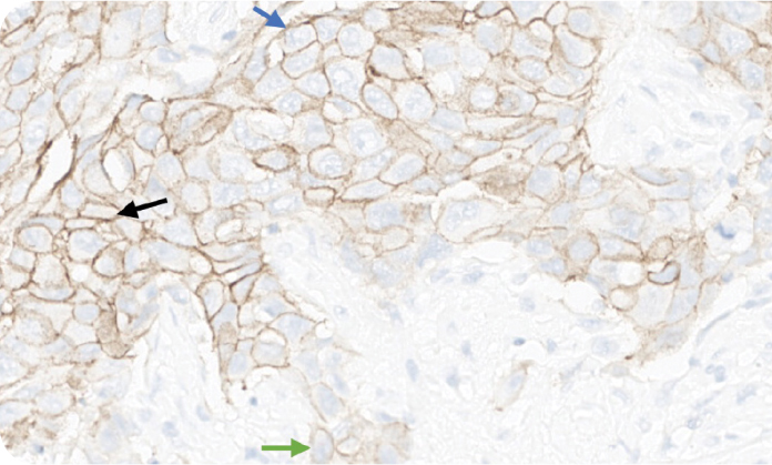
Heterogeneous staining on tumor samples characterized by presence of cells with different levels of HER2 staining can make HER2 IHC interpretation challenging.23,24
Heterogeneous staining, IHC 3+ spanning a range of patterns and intensities from strong complete (25% black arrow) to moderate complete (50% blue arrow) and weak incomplete (20% green arrow).
Microscopic image displaying heterogeneous staining during HER2 interpretation.Microscopic image displaying heterogeneous staining during HER2 interpretation.

Images contributed from HER2Know.com.19

Access educational resources, clinical cases and tools to assess your HER2 IHC scoring consistency in solid tumors.

Visit HER2Know.com
Microscopic slide showing stained cells that are positive for HER2 using immunohistochemistry (IHC)

ASCO, American Society of Clinical Oncology; CAP, College of American Pathologists; HER2, human epidermal growth factor receptor 2; IHC, immunohistochemistry.

Guidelines icon

CAP Guidelines Help Ensure Standardized HER2 IHC Reporting2

Standardization of assay results relies on adherence to proficiency and accreditation requirements.2 CAP recommends that testing laboratories perform ongoing competency assessment and proficiency tests at least twice a year (with performance requirements starting at 90%). Further recommendations include laboratory accreditation as well as onsite inspection every other year and self-inspection every year.2

How HER2 results are reported can have implications for patient care.25 As many as 50% of pathology reports for breast cancer are missing some elements critical to patient management.1

Checklist icon small

To ensure reliable differentiation of HER2-low expression, record discrete scores on each patient’s HER2 IHC report. Recording staining patterns, such as faint/barely perceptible/incomplete staining, and percentage staining is also important.11

Recommended result reporting

IHC 0

HER2-negative

IHC 1+

HER2-negativea

IHC 2+/ISH-

HER2-negativea

IHC 2+/ISH+

HER2-positive

IHC 3+

HER2-positive

ASCO/CAP breast biomarker reporting template guidance11

aBreast cancers with HER2 IHC score 1+ or HER2 IHC score 2+ and a negative ISH result may be reported as ‘HER2-low’

Cannot be determined or test not performed2

Must report HER2 test result as indeterminate if technical issues prevent one or both tests (IHC and ISH) from being reported as positive, negative or equivocal.

Conditions may include:

  • Inadequate specimen handling
  • Artifacts (crush or edge artifacts) that make interpretation difficult
  • Analytic testing failure

Another specimen should be requested for testing to determine HER2 status. Reason for indeterminate testing should be noted in
a comment in the report.

ASCO/CAP comment recommendation2

Please refer to ‘The Bottom Line’ in the 2023 ASCO/CAP guidelines update for HER2 testing report footnote comment recommendation.

IHC 3+

HER2-positive

IHC 2+/ISH+

HER2-positive

IHC 2+/ISH-

HER2-negativea

CAP breast biomarker reporting template guidance11

aBreast cancers with HER2 IHC score 1+ or HER2 IHC score 2+ and a negative ISH result may be reported as ‘HER2-low’

IHC 1+

HER2-negativea

IHC 0

HER2-negative

Cannot be determined or test not performed2

Must report HER2 test result as indeterminate if technical issues prevent one or both tests (IHC and ISH) from being reported as positive, negative or equivocal.

Conditions may include:

  • Inadequate specimen handling
  • Artifacts (crush or edge artifacts) that make interpretation difficult
  • Analytic testing failure

Another specimen should be requested for testing to determine HER2 status. Reason for indeterminate testing should be noted in a comment in the report.

ASCO/CAP comment recommendation2

Please refer to ‘The Bottom Line’ in the 2023 ASCO/CAP guidelines update for HER2 testing report footnote comment recommendation.

Checklist icon

CAP has developed pathology reporting templates to promote complete and standardized reporting of breast cancer specimens for HER2.11 The National Comprehensive Cancer Network® (NCCN®) Breast Cancer Panel endorses the use of CAP protocols for reporting the pathologic analysis of all breast cancer specimens.1

aBreast cancers with HER2 IHC score 1+ or HER2 IHC score 2+ and a negative ISH result are eligible for clinically appropriate HER2-targeted therapy and may be reported as HER2-low.

ASCO, American Society of Clinical Oncology; CAP, College of American Pathologists; HER2, human epidermal growth factor receptor 2; IHC, immunohistochemistry; ISH, in situ hybridization; NCCN, National Comprehensive Cancer Network.

Light key takeaway icon

Find out how to improve HER2 IHC scoring to ensure reliable differentiation of low levels of HER2 expression

Learn more about identifying HER2-low in breast cancer and assigning a HER2 IHC score when encountering challenging staining patterns.

Visit HER2Know.com

HER2, human epidermal growth factor receptor 2; IHC, immunohistochemistry.

Dark key takeaway icon

Considerations for Your Practice

HER2 testing has become routine practice to ensure all patients with breast cancer receive more informed treatment options. Reliable differentiation of scores at the low end of the spectrum may be clinically meaningful for patients with breast cancer.1,2

  • Follow ASCO/CAP best practices for HER2 testing; examine HER2 IHC at high power magnification (40x) when discriminating IHC 0 from 1+ staining; IHC with reflex ISH for 2+ specimens is the recommended methodology2
  • Score consistently across the full spectrum of HER2 expression; in lower levels of HER2 expression, differentiating between HER2 IHC 0 and 1+ supports active research and refinement of testing criteria2
  • Use the CAP reporting template to ensure HER2 test results are in an ideal format for interpretation by the MDT11

ASCO, American Society of Clinical Oncology; CAP, College of American Pathologists; HER2, human epidermal growth factor receptor 2; IHC, immunohistochemistry; ISH, in situ hybridization; MDT, multidisciplinary team.

Helpful Resources

Check out these featured resources on HER2 biomarker testing in breast cancer.

Playlist icon

Add your preferred resources to My Favorites—a personalized list of resources to download and share with your peers or patients.

View My Favorites

dummy

View more resources 

ASCO, American Society of Clinical Oncology; CAP, College of American Pathologists; HER2, human epidermal growth factor receptor 2; IHC, immunohistochemistry.

Email icon

Subscribe to Emails

Subscribe and stay up to date with biomarker testing in breast cancer.

Subscribe

Chat box icon

Connect With Us

Connect with the Precision Medicine Team for actionable support with HER2 testing in breast cancer.

Connect with us

HER2, human epidermal growth factor receptor 2.

  1. Referenced with permission from the NCCN Clinical Practice Guidelines in Oncology (NCCN Guidelines®) for Breast Cancer V.6.2024. © National Comprehensive Cancer Network, Inc. 2024. All rights reserved. Accessed November 20, 2024. To view the most recent and complete version of the guideline, go online to NCCN.org
  2. Wolff AC, et al. J Clin Oncol. 2023;41(22):3867–3872.
  3. Wolff AC, et al. J Clin Oncol. 2018;36:2105–2122.
  4. Tarantino P, et al. J Clin Oncol. 2020;38(17):1951–1962.
  5. Guiterrez C and Schiff R. Arch Pathol Lab Med. 2011;135(11):55–62.
  6. American Cancer Society, Inc., Surveillance Research Breast Cancer Facts & Figures 2019-2020. Accessed August 13, 2024. https://​www.​cancer​.org/​content/​dam/​cancer-org/​research/​cancer-facts​-and-statistics/breast-cancer-facts-and-figures/breast-cancer-facts-and-figures-2019-2020.pdf
  7. Perez EA, et al. Cancer Treat Rev. 2014;40(2):276–284.
  8. Schettini F, et al. NPJ Breast Cancer. 2021;7(1):32.
  9. Rakha EA, et al. J Clin Pathol. 2015;68:93–99.
  10. Modi S, et al. J Clin Oncol. 2020;38(17):1887–1896.
  11. CAP. Template for Reporting Results of Biomarker Testing of Specimens from Patients with Carcinoma of the Breast. Version 1.5.0.1. Accessed August 13, 2024. https://documents.cap.org/documents/Breast.Bmk_1.5.0.1.REL_CAPCP.pdf
  12. US Food and Drug Administration. List of Cleared or Approved Companion Diagnostic Devices (In Vitro and Imaging Tools). Accessed August 13, 2024. https://​www.​fda.​gov/​medical-devices/​in-vitro-diagnostics/​list​-cleared​-or-approved​-companion-diagnostic-devices-in-vitro-and-imaging-tools
  13. Wolff AC, et al. Arch Pathol Lab Med. 2023;147(9):993–1000.
  14. Leica Biosystems. Bond™ Oracle™ HER2 IHC System Instructions For Use. Accessed August 13, 2024. https://​www.​leicabiosystems​.com/​sites/​default/​files/2020-10/Bond_Oracle_HER2_IHC_system_TA9145_ROW-EN-CE_Rev_B.pdf
  15. Bond™ Oracle™ HER2 IHC System Interpretation Guide – Breast Tissue. Accessed August 13, 2024. https://www.leicabiosystems.com/sites/default/files/2020-10/Oracle%20HER2%20Interpretation%20Guide%20%28Breast%29.pdf
  16. Agilent. HercepTest™ for Automated Link Platforms. Package Insert. Accessed August 13, 2024. https://www.agilent.com/cs/library/packageinsert/public/PD04084US_03.pdf
  17. FDA. InSite™ Her-2/neu Package Insert. Accessed August 13, 2024. https://www.accessdata.fda.gov/cdrh_docs/pdf4/p040030c.pdf
  18. PATHWAY anti-HER-2/neu (4B5) Rabbit Monoclonal Primary Antibody. Package Insert. Accessed August 13, 2024. https://​www.​thelancet.​com/​cms/​10.1016/​j.ebiom.2023.​104571/attachment/44694de5-dcf7-4192-90fe-e88e659c3868/mmc2.pdf
  19. Daiichi Sankyo, Inc. and AstraZeneca. Her2Know.com. Accessed August 13, 2024. https://www.her2know.com/
  20. Lai HZ, et al. Cancers (Basel). 2022;14(15):3774.
  21. Rakha EA, et al. J Clin Pathol. 2023;76(4):217–227.
  22. Hicks DG and Schiffhauer L. Lab Med. 2011;42(8):459–466.
  23. Grassini D, et al. Pathobiology. 2022;89:278–296.
  24. Marchiò C, et al. Semin Cancer Biol. 2021;72:123–135.
  25. Vallacha A. Asian Pac J Cancer Prev. 2018;19(3):853–858.

NCCN makes no warranties of any kind whatsoever regarding their content, use or application and disclaims any responsibility for their application or use in any way.

AstraZenecaYoutube

This information is intended for US healthcare professionals only.

Connect With Us Subscribe to Emails Sitemap
Privacy Notice Legal StatementAstraZeneca US Corporate Site Contact Us
Cookie Policy Cookie Notice Consumer Health Data Privacy Notice Your Privacy ChoicesYour Privacy ChoicesYour Privacy Choices

©2025 AstraZeneca. All rights reserved. US-105746 Last Updated 12/25

Share Resource

Share your precision medicine resource(s) via social media or email.

Share to Apps

{{$.copyrighttext}}

©2024 AstraZeneca. All rights reserved. US-86234 Last Updated 11/24

  • Share via  LinkedIn
  • Share via  Facebook
  • Share via  X
  • Share via  Doximity
Share via Email
To add multiple email addresses, please separate them with a comma (e.g. email1@domain.com, email2@domain.com)

errorMessage

requestToSignInContent

Welcome to

AZPrecisionMed.com

This website is intended to help healthcare professionals practicing in the US find and access scientifically balanced and evidence-based information and resources on oncology biomarker testing in precision medicine.

The dissemination of this information may be subject to different medical and regulatory requirements in other countries.

This information is provided for educational purposes only. It is not intended for use in the diagnosis or treatment of individual patients.

Are you a healthcare professional in the United States?

No, take me to AstraZeneca-us.com

What is an AstraZeneca authorised person?

This website is only intended for healthcare professionals practicing in the United States and authorized AstraZeneca company persons. US Headquarters personnel, US Field Diagnostics and US Field Medical personnel are authorized to browse and search the database for their own background knowledge and insight. This website is not intended for use by US Field Sales personnel. None of the content on this site should be directly linked or distributed without approval.

©2024 AstraZeneca. All rights reserved. US-86234 Last Updated 11/24

  • Share via  LinkedIn
  • Share via  Facebook
  • Share via  X
  • Share via  Doximity
Share via Email
To add multiple email addresses, please separate them with a comma (e.g. email1@domain.com, email2@domain.com)

errorMessage

requestToSignInContent

Share Resource

Share your precision medicine resource(s) via social media or email.

Share to Apps

©2024 AstraZeneca. All rights reserved. US-86234 Last Updated 11/24